
Published on July 14, 2025 9:20 PM GMT
Back in Part 1, I found an interesting candidate for a cross-sensory region of the brain (aka used for sight, hearing, touch, etc, alike) that might be active when we consciously perceive sensations and inactive when we only do some kind of subconscious sensory processing.
Both V2 and V5 project back down to the pulvinar nucleus in the thalamus, which is a huge hub for multisensory processing (auditory and tactile as well as visual). The pulvinar is responsible for attentional control, and lesions to it can cause hemispatial neglect (the phenomenon where, often after stroke, a patient might be wholly unaware of a whole half of their body or any sensations coming from that side.)
A summary of my current working model of conscious sensory perception is:
There’s lots of sense data that the brain receives, processes, and uses to guide some behavior, but which we don’t subjectively, consciously perceive.
blindsight, numb touch, deaf hearing: subconscious processing of residual sense data in people with sensory disabilities
subthreshold sensations: subconscious processing of stimuli too faint or ambiguous for us to consciously register
inattentional blindness: subconscious processing of stimuli we aren’t consciously aware of because our attention is otherwise occupied
Conscious sensory perception, compared to unconscious sensory processing, correlates with greater activity (by various metrics) in some brain sensory processing regions (V2 and V5 for sight, the temporal gyrus for hearing, S2 for touch).
Interestingly, in all these cases it’s not the “primary” sensory processing region, i.e. the first to receive inputs from the sensory nerves, that is specific to conscious perception, but rather regions devoted to more complex sensory pattern recognition.
Clearly, since these are different regions for different senses, they don’t constitute a single necessary-and-sufficient neural correlate of conscious sensory perception. There might still be one, though; we gotta keep looking.
So, is it activity in the pulvinar nucleus?
Well, it’s hard to tell for sure.
People (and monkeys) can have damage to a pulvinar nucleus and be almost completely normal in their capacity for everyday life activities, which doesn’t fit well with the idea that the pulvinar is necessary-and-sufficient for conscious sensory perception. On the other hand, maybe the damage in those cases is incomplete. We don’t have a lot of examples of complete bilateral destruction of both pulvinar nuclei.
But there is some suggestive evidence that the pulvinar is at least involved in attention and conscious awareness.
I don’t have a really clear-cut conclusion here, unfortunately, but I’m posting it anyway for the sake of transparency about my learning process.
Neuroscientists who are all “you can’t talk about the “function” of “brain regions”, that’s modern-day phrenology!”, feel free to point and giggle…this time.
I still think I believe in the power of neuroanatomy, especially subcortically. But to some extent that’s layman’s bias — I have to gravitate to simple, old methods because if you do some sort of complicated Big Data functional-connectivity wizardry I won’t be able to tell if the whole thing is concocted out of statistical shenanigans. I am limited by what I can understand, and if the best options are out of my reach, so it goes.
At any rate, it looks like the pulvinar is one of the regions where activity looks different between conscious perceptions and their absence…but it’s hard to say more than that.
Pulvinar Anatomy
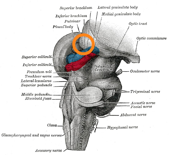
The pulvinar nuclei are clumps of neurons located in the thalamus, at the top of the brainstem. You have one on each side, left and right.
They receive both neuronal input from, and project neuronal output back to:12
the retina
the superior colliculus (another brainstem region and the “first stop” in the brain for visual signals from the retina)
the striate visual cortex, or V1
the extrastriate cortex, or V2-V5
other cortical regions (parietal, temporal, frontal, temporal, cingulate)
the striatum/basal ganglia (a deep structure in the limbic system involved in motor responses and motivation)
the amygdala (also limbic, involved in reactions to threats)
Basically, it’s a major hub. In primates most of it seems to be devoted to vision, but it connects everywhere, especially to the cortex.
Pulvinar Lesions
We can learn a lot about a brain region from lesion studies — what happens to the organism when it’s damaged? (Through disease or injury in humans, or through experimentally destroying it in animals.)
One thing pulvinar stroke lesions do (compared to stroke lesions elsewhere in the brain) is cause spatial neglect. 3
This is the phenomenon where a patient “neglects” a part of the space around them — maybe their left or right side. Someone with left-sided hemispatial neglect, for instance, might not eat the food on the left side of their plate, might not shave or make up the left half of their face, might copy only the right half of a drawing, or might bump into objects on their left side.
So that’s promising for the pulvinar as a site of conscious sensory awareness; patients with spatial neglect are unaware of pretty much every sensory perception on their “bad” side.
But not all pulvinar lesions cause complete hemispatial neglect. Often, one-sided pulvinar lesions seem to cause subtler difficulties in selective attention to stimuli on one side, especially under conditions when the patient is tasked to focus on a “target” stimulus and ignore a “distractor.” This does suggest a role in attention; but the studies on pulvinar lesions are frustratingly inconsistent. Different studies find different deficits; some studies find no deficits at all; generally humans and monkeys with pulvinar lesions have almost normal sensory and motor function, which is not quite what you’d see if the pulvinar was “the” seat of conscious sensory perception.
Subjects with pulvinar lesions may have no obvious symptoms beyond limb weakness, clumsiness, and tactile sensory issues (numbness or pain) on the side of the body opposite to the lesion; they can even make a full recovery with no sensory or motor deficits.4
When mild pulvinar lesion symptoms exist, they’re often a sort of “lesser version of hemispatial neglect”. The neglect patient completely ignores the contralesional side (opposite the lesion); other pulvinar lesion patients merely exhibit an attentional bias to the ipsilesional side (same side as the lesion), or a subtle impairment in difficult sensory-attention tasks on the contralesional side.
For instance, one patient couldn’t visually follow lines from left to right (into his contralesional field). His eyes fixed for longer on a single point than normal controls, and he was worse than controls at visually searching for items on the contralesional side. This is a clearcut example of something like a deficit in visual “exploration” on the bad side.5
People with pulvinar lesions also have impairments related to voluntary control over their eye movements. In a task where participants are instructed to look at a particular colored circle when it appears, their eyes are often drawn unintentionally to a “distractor” circle of a different color. Pulvinar lesion patients (without hemispatial neglect) are worse at the task when the “distractor” is on the opposite side from the lesion (contralesional); healthy controls are better at the task overall and show no “side” preference.6 Similar results happen when it comes to reporting the color of a “target” vs “distractor” square; pulvinar lesions interfere with “response competition”, the ability to respond to the target rather than distractor stimulus.7
One theory of what’s going on is that shifting visual attention involves three operations: “disengage” from what you’re currently looking at, “move” to a new location, and “engage” with the new visual stimulus. The pattern of impairments in pulvinar lesions is consistent with a problem with the “engage” step, in the contralesional visual field. They can move their gaze there — this isn’t true neglect — but they don’t get the boost in speed and accuracy that normal people get when the “spotlight of attention” is focused on a region.8
Monkeys with pulvinar lesions, similarly, have their gaze more strongly drawn to visual stimuli than a blank screen; they get “visually captured” more, and scan less, than control monkeys.9 In tasks with distractors, monkeys do much worse on the contralateral side when their pulvinar is inactivated (with drugs). 10; they also have much less neural response to visual stimuli in V4, and see a large increase in alpha wave power across the whole cortex, a state more similar to sleep.
Lesions in the dorsal pulvinar can also cause deficits in reaching and grasping for things in a human subject.11
Drug-based inactivation of the pulvinar nucleus in monkeys had similar results related to reaching/grasping; monkeys would not use the hand on the non-lesioned side to reach for a treat, and if their “good hand” was inactivated, they were awkward in reaching and grasping with their “bad hand”, sometimes letting the treat fall to the floor altogether (which never happens with intact monkeys.) The monkeys were neither paralyzed nor blind on the contralesional side; they simply did not explore it, either with eyes or limbs, especially if there was something happening simultaneously on the ipsilesional side. They were still capable of directing their gaze towards contralesional images, but their gaze went faster to the ipsilesional side and stayed there.12
On the other hand, some other experimental lesions of the entire pulvinar nucleus in monkeys resulted in no effects on eye movements, so maybe not, after all.13
The “neglect-like” effect of pulvinar lesions isn’t restricted to vision but is relevant to other senses. A pulvinar lesion, in one case, caused an unusual auditory deficit — the patient could hear on both sides if the sound only came from one side at a time, but could not hear anything in his left ear if different sounds were coming from each side simultaneously, as though the problem was in attending to left-sided sounds in the presence of competing right-sided sounds.14
What happens if you have damage to both pulvinar nuclei? In one case, sudden-onset double vision and nystagmus (wobble) when instructed to move his eyes.15
The Pulvinar and Blindsight
Monkeys with blindsight have damage to the primary visual cortex V1, but can still be trained to move their eyes accurately to targets in their “blind field.”
If you also inactivate the pulvinar nucleus by injecting a compound that blocks neuron firing, the monkeys no longer show evidence of blindsight.
By contrast, the lateral geniculate nucleus (LGN) is not necessary for blindsight; it usually deteriorates in response to V1 damage, so it’s mostly gone already even in animals that still have blindsight.
Now, remember that blindsight is (usually) a form of unconscious visual processing — except in cases that are arguably a sort of “consciousness without visual qualia”, where the subject reports knowing about or sensing visual stimuli but not truly “seeing” them.
If the pulvinar is active in blindsight in monkeys, that could mean pulvinar activity is not sufficient for making a visual perception conscious; or the pulvinar could be involved in this sort of “technically conscious blindsight”. The monkey can’t tell us what it’s experiencing! So from animal experiments alone, We Just Don’t Know.16
The Pulvinar and Confidence
If you give a monkey a visual categorization task, with the option to “opt out” if uncertain, there’s less activity in the monkey’s pulvinar nucleus when they’re choosing to opt out than when they’re making a choice. In other words, activity in the pulvinar seems to track “subjective confidence” in their perceptions, or at least their willingness to “bet” on their perceptions. If you inactivate the thalamus, the monkey’s performance on the task is not impaired, but it is much less confident (i.e. more likely to choose the “safe” opt-out button.) By contrast, if you inactivate the LGN, the monkey’s performance is impaired.17
This is interestingly reminiscent of blindsight in humans. Not only do humans with blindsight report being unable to see, but if asked to bet on their confidence level on vision tasks, they usually bet as though they expect to perform at chance — even when in fact, they are getting enough subconscious visual data to perform well above chance.
Perhaps “knowing when you’re right” about visual data — a decent proxy for consciousness — requires pulvinar activity.
The Pulvinar and Attention
In a distractor task, where subjects were asked to pay attention either to the top two or bottom two of four images arranged in a square, only the images people pay attention to show spatially localized activity in the pulvinar nucleus (i.e. fMRI-measured activity is concentrated in different parts of the pulvinar depending on whether the image is on the left or right of the screen). The unattended images don’t spatially localize. 18
That is, the spatial pattern of activity in the pulvinar depends on whether you’re paying attention or not. Without attention, activity is more diffuse; with attention, the activity pattern carries spatial information about the image.
The Pulvinar and Arousal
Interestingly, the pulvinar nucleus is much less active than other parts of the thalamus during anaesthesia, which is suggestive of a role in consciousness.19
Also tantalizing: when a macaque’s pulvinar is inactivated, it gains a lot of power in the alpha EEG frequency throughout the cortex, and diffuse activation of visual-cortex neurons that are normally only active in the presence of particular visual stimuli. This is a pattern ordinarily seen in sleep.20
One patient developed severe hypersomnia — sleeping almost continuously, all day long — after a stroke that damaged the right thalamus, including the pulvinar nucleus on that side.21
The Pulvinar and Conscious Perception
You can train a monkey to report whether it sees a visual stimulus or not, including during optical illusions where the presence of distractors can make a target seem to suddenly “disappear.”
If you do this while recording neural responses, some visual processing areas (e.g. the LGN) show the same pattern of response whether the monkey reports seeing the target or not; but the pulvinar shows quite different responses, with some neurons being more responsive to “invisible” trials and some neurons being more responsive to “visible” ones. The effect in the pulvinar is bigger than that in visual cortex areas like V1, V2, and V4 using the same method.22
If you then don’t have the monkey press the button, the results are the same; pulvinar neural responses differ between trials where the monkey “should” be able to see the target and trials where it “shouldn’t.” This means it’s not simply tracking something like the hand movement or the decision to move.
Conclusions
The pulvinar is one of the regions (but not the only one) where lesions can lead to hemispatial neglect, or the total lack of conscious attention/perception on one side of the body. But damage or inhibition of the pulvinar usually doesn’t do anything so drastic, which suggests that there are multiple brain regions sufficient for perceptual attention.
The pulvinar definitely changes its activity pattern based on “consciousness-related” states like attention, arousal, and confidence in one’s correctness on perceptual tasks.
Mess with the pulvinar and you can (but don’t always) get phenomena like:
impaired performance on visual tasks on the affected “side”
lower confidence in visual tasks even when performance is not impaired
impairments in other sensorimotor tasks (auditory processing, reaching/grasping), again on the affected side
loss of blindsight when it was previously present
excessive sleep, and sleep-like brainwave patterns
So, while this isn’t exactly the smoking gun I initially hoped it was, I do think the pulvinar is (weasel word alert) “involved in” conscious perception.
Zhou, N. A., et al. "The mouse pulvinar nucleus: Organization of the tectorecipient zones." Visual neuroscience 34 (2017): E011.
Grieve, Kenneth L., Carlos Acuña, and Javier Cudeiro. "The primate pulvinar nuclei: vision and action." Trends in neurosciences 23.1 (2000): 35-39.
Karnath, Hans‐Otto, Marc Himmelbach, and Chris Rorden. "The subcortical anatomy of human spatial neglect: putamen, caudate nucleus and pulvinar." Brain 125.2 (2002): 350-360.
Van der Stigchel, S., et al. "Oculomotor integration in patients with a pulvinar lesion." Neuropsychologia 48.12 (2010): 3497-3504.
Ogren, Marilee P., Catherine A. Mateer, and Allen R. Wyler. "Alterations in visually related eye movements following left pulvinar damage in man." Neuropsychologia 22.2 (1984): 187-196.
Van der Stigchel et al (2010).
Danziger, Shai, et al. "Contributions of the human pulvinar to linking vision and action." Cognitive, Affective, & Behavioral Neuroscience 4.1 (2004): 89-99.
Rafal, Robert D., and Michael I. Posner. "Deficits in human visual spatial attention following thalamic lesions." Proceedings of the National Academy of Sciences 84.20 (1987): 7349-7353.
Ungerleider, Leslie G., and Carol A. Christensen. "Pulvinar lesions in monkeys produce abnormal eye movements during visual discrimination training." Brain Res 136.1 (1977): 189-196.
Zhou, Huihui, Robert John Schafer, and Robert Desimone. "Pulvinar-cortex interactions in vision and attention." Neuron 89.1 (2016): 209-220.
Wilke, Melanie, et al. "Reach and grasp deficits following damage to the dorsal pulvinar." cortex 99 (2018): 135-149.
Wilke, Melanie, et al. "Pulvinar inactivation disrupts selection of movement plans." Journal of Neuroscience 30.25 (2010): 8650-8659.
Bender, D. B., and J. S. Baizer. "Saccadic eye movements following kainic acid lesions of the pulvinar in monkeys." Experimental brain research 79 (1990): 467-478.
Hugdahl, Kenneth, Knut Wester, and Arve Asbjørnsen. "Auditory neglect after right frontal lobe and right pulvinar thalamic lesions." Brain and Language 41.3 (1991): 465-473.
Ohtsuka, Kenji, et al. "Pursuit deficits in bilateral pulvinar lesions." Ophthalmologica 203.4 (1991): 196-202.
Kinoshita, Masaharu, et al. "Dissecting the circuit for blindsight to reveal the critical role of pulvinar and superior colliculus." Nature communications 10.1 (2019): 135.
Komura, Yutaka, et al. "Responses of pulvinar neurons reflect a subject's confidence in visual categorization." Nature neuroscience 16.6 (2013): 749-755.
Fischer, Jason, and David Whitney. "Attention gates visual coding in the human pulvinar." Nature communications 3.1 (2012): 1051.
Zhou et al (2017).
Zhou et al (2016).
Hansen, Peter Nørregaard, et al. "Severe hypersomnia after unilateral infarction in the pulvinar nucleus–a case report." BMC neurology 20.1 (2020): 442.
Wilke, Melanie, Kai-Markus Mueller, and David A. Leopold. "Neural activity in the visual thalamus reflects perceptual suppression." Proceedings of the National Academy of Sciences 106.23 (2009): 9465-9470.
Discuss
