


Deep Visual Proteomics: Integrating AI and Mass Spectrometry for Cellular Phenotyping:
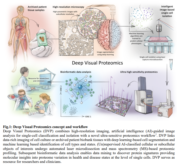
Deep Visual Proteomics (DVP) revolutionizes the analysis of cellular phenotypes by combining advanced microscopy, AI, and ultra-sensitive mass spectrometry (MS). Traditional methods often target a limited subset of proteins, but DVP extends this capability by enabling comprehensive proteomic analysis within the native spatial context of cells. This approach involves high-resolution imaging for single-cell phenotyping, AI-driven cell segmentation, and automated laser microdissection to isolate cellular or subcellular regions of interest precisely. These isolated samples are subjected to ultra-high sensitivity mass spectrometry for detailed proteomic profiling.
Developed using the ‘BIAS’ (Biology Image Analysis Software), DVP facilitates seamless integration of imaging and proteomic technologies. It enables the identification of distinct cell types and states based on AI-defined features, enhancing the accuracy and efficiency of cellular phenotyping. Applications of DVP span from studying single-cell heterogeneity to characterizing proteomic differences in disease tissues like melanoma and salivary gland carcinoma. By preserving spatial information alongside molecular insights, DVP offers a powerful tool for advancing research and clinical diagnostics in cell and disease biology.
Image Processing and Single Cell Isolation Workflow in Deep Visual Proteomics:
The image processing and single-cell isolation workflow in DVP integrates cutting-edge microscopy technologies with advanced AI-driven image analysis and automated laser microdissection. Beginning with high-resolution scanning microscopy, the process involves capturing whole-slide images that are processed using the BIAS. BIAS supports various microscopy formats and utilizes deep learning algorithms to segment cellular components like nuclei and cytoplasm precisely. This includes innovative techniques like image style transfer to optimize deep learning model training for specific biological contexts. BIAS facilitates seamless interaction with laser microdissection systems such as ZEISS PALM MicroBeam and Leica LMD6 & 7, ensuring accurate transfer and automated targeted cell extraction. This integrated workflow enables rapid and precise single-cell isolation, which is crucial for in-depth proteomic analysis of cellular and tissue samples in DVP applications.
Characterizing Single Cell Heterogeneity with Deep Visual Proteomics:
DVP enables the characterization of functional differences among phenotypically distinct cells at the subcellular level. Applying this workflow to an unperturbed cancer cell line, researchers used deep learning-based segmentation to isolate and analyze individual cells and nuclei. This approach addressed the challenges of processing minute samples, allowing direct analysis from 384 wells using advanced mass spectrometry. The proteomic profiles of whole cells and isolated nuclei were distinct, with high reproducibility. Machine learning identified six classes of nuclei with significant morphological and proteomic differences. This demonstrated that visible cellular phenotypes correspond to distinct proteome profiles, offering insights into cell cycle regulation and potential cancer prognostic markers.
DVP Uncovers Cancer Tissue Heterogeneity:
DVP offers high-resolution, unbiased proteomic profiling of distinct cell classes within their spatial environments. Applied to archived salivary gland acinic cell carcinoma tissue, DVP revealed significant proteomic differences between normal and cancerous cells. Normal acinar cells showed high expression of secretory proteins, while cancer cells exhibited elevated interferon-response proteins and the proto-oncogene SRC. Extending this to melanoma, DVP differentiated central tumor cells from those at the tumor-stroma border, identifying distinct proteomic signatures linked to disease progression and prognosis. These findings underscore DVP’s potential for precise molecular disease subtyping, guiding clinical decision-making.
Outlook for DVP:
The DVP pipeline integrates high-resolution microscopy with advanced image recognition, automated laser microdissection, and ultra-sensitive MS-based proteomics. This robust system applies to diverse biological systems that can be microscopically imaged, from cell cultures to pathology samples. DVP allows the rapid scanning of slides to isolate rare cell states and study the extracellular matrix’s proteomic composition. With the potential for super-resolution microscopy, DVP can achieve precise cell state classification. By combining powerful imaging technologies with unbiased proteomics, DVP offers significant applications in basic biology and biomedicine, particularly in oncology, where it enhances digital pathology by providing a comprehensive proteomic context.
Check out the Paper. All credit for this research goes to the researchers of this project. Also, don’t forget to follow us on Twitter.
Join our Telegram Channel and LinkedIn Group.
If you like our work, you will love our newsletter..
Don’t Forget to join our 46k+ ML SubReddit
The post Revolutionizing Cellular Analysis: Deep Visual Proteomics Integrates AI and Mass Spectrometry for Advanced Phenotyping appeared first on MarkTechPost.
