


Aging is linked to a significant rise in neurodegenerative diseases like Alzheimer’s and cognitive decline. While brain aging involves complex molecular and cellular changes, our understanding of these processes within their spatial context remains limited. Past studies have provided valuable insights into age-related brain changes at a single-cell level but lack comprehensive spatiotemporal resolution. High-throughput spatial omics offer the potential for uncovering cell interactions during aging, yet current research focuses either on spatial or temporal aspects, not both. Advanced computational tools are urgently needed to analyze spatial omics data, enabling a deeper understanding of cell-type-specific changes and interactions during aging.
Stanford University and UCLA researchers created a spatially resolved single-cell transcriptomics atlas of 4.2 million mouse brain cells spanning 20 age points across the adult lifespan. They also examined the effects of rejuvenating interventions, such as exercise and partial reprogramming. They developed spatial aging clocks using this atlas—machine learning models identifying transcriptomic aging patterns, rejuvenation, and disease. Their findings reveal that T cells have a pro-aging effect on nearby cells, while neural stem cells exert a rejuvenating influence. These insights highlight the significant impact of rare cell types on brain aging and offer potential targets for anti-aging therapies.
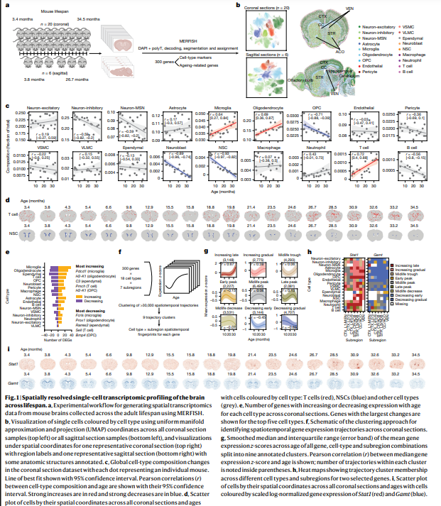
The study constructed a comprehensive single-cell transcriptomic atlas of the mouse brain, profiling 4.2 million cells across 20 age points spanning adulthood. This allowed for the creating of spatial aging clocks—machine learning models trained to identify aging, rejuvenation, and disease-related transcriptomic signatures across different brain regions and cell types. The method also considered rare cell populations, enhancing the precision of age-related changes in the brain. By leveraging these spatial clocks, the researchers were able to detect cell-type-specific patterns linked to aging processes, providing a detailed understanding of age-related shifts in brain biology.
In addition, deep learning methods were used to see the role of specific cell types in aging and rejuvenation. The study revealed that T cells infiltrate the brain with age and have a pro-aging impact on neighboring cells, while neural stem cells exert rejuvenating effects on surrounding tissue. These findings were linked to specific molecular mediators, suggesting that targeting certain cell types might effectively combat tissue aging. This highlights the potential for therapeutic strategies to modulate cell interactions in aging brains to promote rejuvenation and reduce age-related decline.
The study developed spatial transcriptomic clocks by analyzing 4.2 million cells across 20 distinct ages in the adult mouse brain. These clocks identified spatial and cell-type-specific transcriptomic signatures associated with aging, rejuvenation, and disease, including those of rare cell types. Notably, T cells, which increase in the brain with age, were found to have a pro-aging effect on neighboring cells. Conversely, neural stem cells exhibited a pro-rejuvenating impact in adjacent cells. The research also identified potential mediators of these effects, offering insights into cellular interactions that influence brain aging.
In conclusion, the study provides a detailed spatial analysis of aging in the mouse brain, enabling the tracking of gene expression changes across regions and cell types. The developed spatial aging clocks can be used to assess the effects of interventions on aging and disease processes at single-cell resolution. The authors highlight the need for further research to understand the mechanisms behind cell proximity effects, particularly in neurons. They suggest that more in-depth studies, including functional assays and deeper imaging, are required to fully elucidate how T cells and neural stem cells influence brain aging and potential therapeutic strategies for enhancing resilience during aging.
Check out the Paper. All credit for this research goes to the researchers of this project. Also, don’t forget to follow us on Twitter and join our Telegram Channel and LinkedIn Group. Don’t Forget to join our 60k+ ML SubReddit.
 Trending: LG AI Research Releases EXAONE 3.5: Three Open-Source Bilingual Frontier AI-level Models Delivering Unmatched Instruction Following and Long Context Understanding for Global Leadership in Generative AI Excellence….
The post Researchers at Stanford Use AI and Spatial Transcriptomics to Discover What Makes Some Cells Age Faster/Slower in the Brain appeared first on MarkTechPost.
